
Consider a shopkeeper in his mid-fifties near Rajpura, on the outskirts of Patiala, who has lived with type 2 diabetes for close to nine years. By any reasonable standard, he is a careful patient. His HbA1c is checked on schedule. His feet are examined at every diabetes review. Ask him, though, when he last sat through a dilated retinal examination — not the quick vision chart at the optician’s, but an actual dilated look inside the eye — and he genuinely cannot recall. No one along the way ever told him it mattered. And by his own account, his eyesight is perfectly fine.

That last detail is where the real danger sits. A patient can feel that their vision is fine while the retina behind that vision is already bleeding — the two are not connected the way most people assume.

This illustrative scenario repeats itself across the Malwa belt more often than most diabetic patients realise. People with diabetes tend to be disciplined about the parts of the disease that come with a number attached to them — blood sugar, kidney function, cholesterol. Very few extend that same discipline to an annual eye check, largely because the eye gives no early warning the way a foot ulcer does. Closing that gap, between how seriously patients guard the rest of their body and how casually they treat their eyes, is the entire purpose of this article.

What Diabetic Retinopathy Actually Is

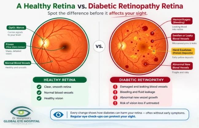
Diabetic retinopathy refers to progressive damage to the small blood vessels of the retina, caused by years of elevated blood sugar. It advances through recognised stages: mild, moderate and severe non-proliferative retinopathy (NPDR), followed by proliferative diabetic retinopathy (PDR), in which abnormal, fragile new vessels begin forming on the retina’s surface. A related complication, diabetic macular oedema (DME), involves fluid accumulation in the macula — the retina’s central, sharp-focus zone — and can appear at any stage of the disease.
What Diabetic Retinopathy

The mechanism itself is easier to describe than the disease is to detect: sustained high blood sugar weakens the walls of the retina’s finest blood vessels over years. They begin leaking, forming microaneurysms, and eventually become blocked, cutting off blood supply to sections of the retina. Starved of oxygen, the eye responds by sprouting new, structurally weak vessels — and it is precisely at this stage that the risk of sudden, severe vision loss climbs sharply.

Why Normal Vision Doesn't Mean a Normal Retina

This is arguably the single most important idea in this piece: what a patient reports feeling and what a dilated exam or optical coherence tomography (OCT) scan actually reveals are two separate things. Mild-to-moderate retinopathy very often produces no symptoms whatsoever. Someone can read small print, drive comfortably after dark, and pass an informal vision check while microaneurysms and early haemorrhages are already sitting on their retina. Subjective vision was never designed to be a screening tool. A dilated examination or retinal imaging is the only test that actually qualifies as one.

Why the Screening Clock Matters More Than the Treatment Menu

Early retinopathy generally responds well to monitoring and tighter control of the underlying risk factors. Advanced, unmonitored disease, on the other hand, frequently causes vision loss that no treatment can fully undo. That asymmetry — early intervention works reliably, late intervention often cannot fully compensate — is why how often a patient is screened tends to matter more for long-term vision than which treatment technology eventually gets used.

Inside a Diabetic Retinopathy Screening Visit

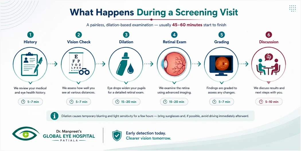
A large part of why diabetic patients put this exam off is simply not knowing what to expect. It runs longer than a routine eye test, but nothing about it is painful, and the sequence is consistent from patient to patient.
Inside a Diabetic Retinopathy Screening

The appointment opens with a history — how long diabetes has been present, how glycaemic control has trended — followed by a visual acuity check. Dilating drops are then administered, taking roughly 20 to 30 minutes to reach full effect, and causing temporary blurring and light sensitivity for a few hours afterward; carrying sunglasses and arranging for someone else to drive is worth planning ahead for. With the pupils dilated, the retina is examined directly, frequently backed up by fundus photography or OCT imaging for precise documentation. Any retinopathy found is graded by severity, and the visit closes with a clear conversation about the finding and what comes next — a routine annual recall, or a referral for closer monitoring or treatment.

Who Actually Needs Screening, and On What Timeline

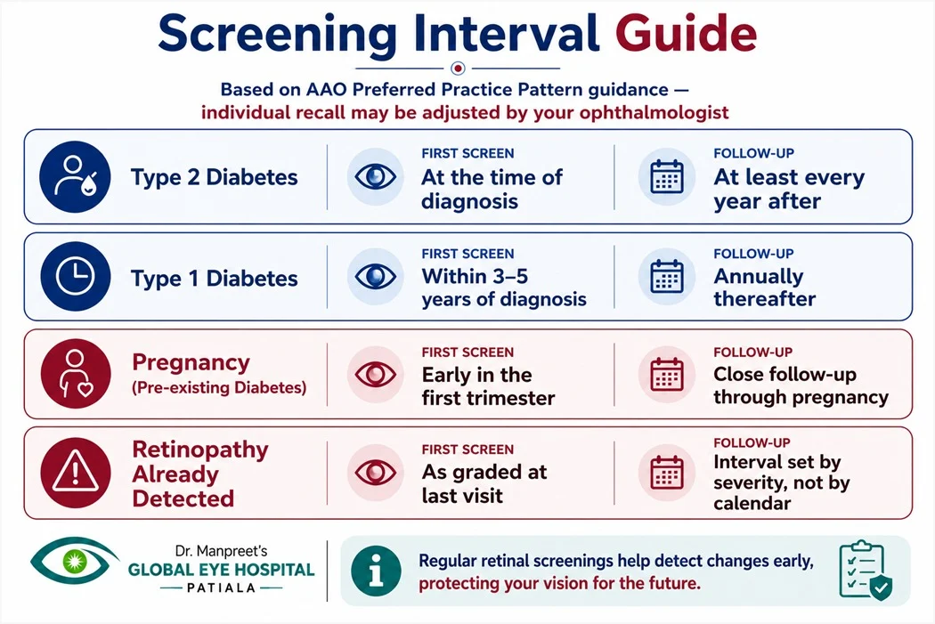
This guidance is deliberately specific rather than a vague “get checked sometimes” suggestion — and none of it changes based on how good a patient’s vision currently feels.
Diabetic Retinopathy guide

Per the American Academy of Ophthalmology’s Diabetic Retinopathy Preferred Practice Pattern, patients with type 2 diabetes should undergo a first retinal screening at the point of diagnosis, then at least once every year afterward. Patients with type 1 diabetes should begin screening within three to five years of diagnosis, continuing annually from there. Pregnant women who already have type 1 or type 2 diabetes should be screened early in the first trimester, with tighter follow-up through the pregnancy, since pregnancy itself can speed up retinopathy progression. Once retinopathy has actually been found, the follow-up schedule is set by how advanced it is, not by a fixed yearly calendar — tighter review is warranted where control is poor, where the disease is progressing, or where macular oedema is present.

Two further points deserve to be stated without softening. First, risk rises sharply with how long diabetes has been present: published data shows that roughly 60% of patients will show some degree of retinopathy after ten years of diabetes, climbing to close to 80% by the fifteen-year mark. Second — and this is the more uncomfortable one — the American Academy of Ophthalmology notes that only around 60% of people with diabetes actually receive the yearly screening they are supposed to. A meaningful share of at-risk patients, in other words, simply are not being examined closely enough, or often enough.

“My vision feels fine, so this year can wait” is, in nearly every case, exactly the reasoning that allows silent progression to continue unchallenged.

Why Diabetic Retinopathy Deserves More Attention in Punjab Than It Gets

India’s diabetic population is projected to reach approximately 79.4 million by 2030. Among people already living with diabetes nationally, an estimated 34.6% will go on to develop some form of diabetic retinopathy, and 10.2% will develop a vision-threatening form of the disease — figures drawn from published ophthalmology research, not from any hospital’s own marketing material.

The 79.4 million / 34.6% / 10.2% figures above are drawn from a study published in the Indian Journal of Ophthalmology. Re-verify the exact citation and figures against the primary source before this article is published, and include the formal citation in the final piece.

What matters at a state level is not only the scale of India’s diabetes burden, but a documented, specific gap between diagnosis and actual eye screening. A qualitative research study examining barriers to diabetic retinopathy screening within public health facilities in Punjab and Chandigarh — built on interviews with people living with diabetes alongside retina specialists, ophthalmologists, optometrists, medical officers, and community health workers — found that low awareness, weak referral pathways from general diabetes care into ophthalmology, and rural-urban access gaps together keep diabetics unscreened for years, despite an already-confirmed diagnosis.

The Punjab/Chandigarh screening-barriers study cited above confirms that this research exists and covers the state broadly; it does not produce a Patiala-specific or hospital-specific prevalence figure, and none should be invented. Source any Patiala-level statistic directly before adding it.

The practical implication for Patiala and the surrounding Malwa belt is straightforward: a meaningful share of the region’s diabetic population has very likely gone years without a dilated retinal exam — not out of carelessness, but because nothing in their regular diabetes care routine ever pointed them toward one clearly. A structured, genuinely accessible diabetic retinopathy screening programme in Patiala answers a documented state-wide gap directly, rather than functioning as one more generic service listing on a hospital website.

If Screening Finds Something, Here Is What Treatment Looks Like

Detecting retinopathy is not, on its own, an emergency — the response is calibrated precisely to the stage found, not applied as one uniform reaction to a single diagnosis.

Retinopath
Mild NPDR is generally managed through monitoring, with priority given to tightening blood sugar and blood pressure control, alongside a shorter recall interval. Moderate-to-severe NPDR calls for closer monitoring, and depending on specific findings, may justify earlier intervention. Proliferative diabetic retinopathy (PDR) is typically managed with anti-VEGF intravitreal injections, panretinal photocoagulation (PRP) laser, or both, aimed at halting further growth of abnormal vessels. Diabetic macular oedema, wherever it is present, is most often treated with anti-VEGF therapy as the first-line approach. In advanced or complicated cases — vitreous haemorrhage, for example — vitrectomy surgery may become necessary.

What treatment can promise, and what it cannot !!

Treatment can frequently slow or halt further progression and preserve remaining vision — but it does not reliably restore vision already lost to advanced, untreated disease. This is exactly why catching the disease early carries more weight than any single treatment technology available today.

Screening and Treatment are not the Same Job

Patients tend to use these two words interchangeably, but they describe two distinct clinical activities, aimed at two different groups of people entirely.

dr manpreet global eye hospital
Screening Treatment
Purpose Detect disease before symptoms appear Manage disease that has already been detected
Who needs it Every diabetic, type 1 or type 2, regardless of current vision Only patients with confirmed retinopathy or macular oedema
Frequency Routine, interval-based (see screening guide above) As clinically indicated by severity and progression
Patient experience Painless, dilation-based examination Injection, laser, or surgical, depending on severity
Outcome if skipped Silent progression, undetected until advanced Progressive, potentially irreversible vision loss

Why Patients across Patiala, Punjab Choose NABH-accredited Global Eye Hospital Patiala?

Global Eye Hospital, Patiala is an NABH-accredited super-specialty eye hospital under the clinical leadership of Dr Manpreet Singh, Chief Eye Surgeon, with diabetic retinopathy care led directly by Dr Balbir Khan, Retina Surgeon. For diabetic patients across Patiala and the wider Malwa belt — Rajpura, Nabha, Samana, Sirhind, Fatehgarh Sahib, Sangrur and Patran — the value of a dedicated diabetic retinopathy screening programme is practical, not promotional: a documented regional access gap exists, and closing it starts with making screening genuinely easy to reach, easy to understand, and easy to act on.

Global Eye Hospital Patiala

According to Dr Balbir Khan, Retina Surgeon at Global Eye Hospital, Patiala, the patients at greatest risk are frequently the ones who feel the least urgency — those whose sugar control looks acceptable on paper and whose vision, subjectively, has not changed. That is exactly the patient profile in whom retinopathy is most easily missed without a dedicated screening visit.

Preparing for Your First Screening Visit

Bring a record of how long diabetes has been present, recent HbA1c readings where available, and a current medication list — particularly for blood pressure and cholesterol, since both influence retinopathy risk directly. Plan for the visit to take 45 to 60 minutes once dilation is factored in, and arrange transport home if you can, since driving immediately after dilation is not advisable. Above all, treat a normal result as a genuinely useful outcome in its own right — it simply means the next screening is a year away, not that the visit achieved nothing.

Frequently Asked Questions

No. Mild and even moderate retinopathy frequently cause no noticeable symptoms. Normal subjective vision does not mean the retina is unaffected — only a dilated exam or imaging can confirm that.

Blurring and light sensitivity typically last a few hours. Sunglasses help, and avoiding driving immediately afterward is advisable.

No. It involves dilating eye drops and a light-based examination or imaging. Screening itself involves no injection or incision.

For type 2 diabetes, at the time of diagnosis. For type 1 diabetes, within three to five years of diagnosis, per American Academy of Ophthalmology guidance.

Yes. Pregnancy can accelerate retinopathy progression in women with pre-existing diabetes, which is why screening is recommended early in the first trimester with closer follow-up.

Yes. Screening recommendations are based on having diabetes and its duration, not on whether it is managed with tablets, insulin, or diet alone.

Good glycaemic control meaningfully reduces risk but does not eliminate it. Screening remains necessary regardless of how well blood sugar is controlled.

Optical coherence tomography is a quick, non-contact imaging scan producing a detailed cross-sectional view of the retina, often used to detect or monitor macular swelling.

No. Diabetic retinopathy can develop or progress over time, which is why annual screening is recommended even after a normal result.

Early changes are typically managed through closer monitoring and tighter glycaemic and blood pressure control, rather than immediate treatment — the approach depends on the specific grade found.

Treatment can often halt or slow progression and help preserve remaining vision, but it does not reliably reverse vision already lost to advanced disease — which is why early detection matters more than any single treatment technology.

Cost depends on the screening method used, whether treatment is required, and the specific technology involved. The first step is a screening visit to establish what, if anything, is actually present.

Medical Disclaimer

This article is intended for general information and does not replace individual medical advice. Diabetic retinopathy screening needs, findings and treatment recommendations vary by patient and can only be determined through a comprehensive eye examination by a qualified ophthalmologist. If you have diabetes and have not had a recent dilated eye examination, consult an eye care professional to arrange one.